


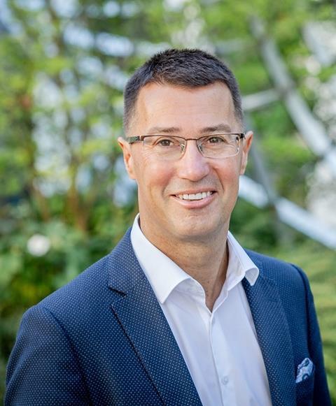
“It is great to come up with wonderful new analytical techniques and use them for clinical questions so that we can advance healthcare,” says Ron Heeren, Distinguished Professor and scientific director at the Maastricht Multimodal Molecular Imaging Institute. He has been dedicated for many years to innovations in mass spectrometry and their applications in various disciplines. In his lecture, he will discuss clinical diagnostics.
Trained as a technical physicist, Heeren and his team developed new physical measurement methods to map the distribution of molecules on complex surfaces (Mass Spectrometry Imaging - MSI). Ten years ago he went to Maastricht to set up a new institute, the Maastricht Multimodal Molecular Imaging Institute, where MSI was further developed in a biomedical setting. In the institute, specialists from various disciplines such as surgeons, mass spectrometrists, mathematicians, pathologists, and electron microscopists work together on solutions for clinical diagnostics. The resulting special research dynamics regularly lead to surprising solutions.
One of the goals is to better understand the complexity of molecular biology in the human body. And to gain new insights into how a certain biological process/disease functions and how we could better intervene with a personal treatment plan (personalized medicine).
Spatial aspect
Heeren emphasizes the importance of the spatial aspect of MSI. “By mapping the natural environment of the cell, you learn which other cells and signals in the biological system the cell has to deal with and can therefore respond to differently. This may be different in different contexts. For example, the metabolism of a liver cell may be altered compared to a normal liver cell due to a nearby tumor.”
Innovations
Heeren and his team have been working for many years on all kinds of MSI innovations that make clinical diagnosis easier and faster, especially for pathologists and surgeons.
“We have worked on the various parts of MSI for this purpose. For example, we have implemented a beam on the source side (ionization source) that can be micrometer-focused to look into the cells. We have improved the detector: if you focus on a very small spot, you generate fewer ions in the mass spectrometer and you therefore need a very sensitive detector. You also want to be able to unravel the ions better in terms of their molecular complexity. To this end, the resolution of the mass spectrometers has been improved over the years.”
Characterization of individual cells
Five years ago, Heeren would not have thought that they would be able to characterize individual cells. “Now we can tell which type of cell we are looking at, for example, a B cell or T cell. We can also identify the metabolic state of the cell: is it an activated or a dormant cell and how old is the cell? This information about a piece of tissue contributes to a better understanding of the local complexity. In addition to MSI, other techniques like optical microscopy, electron microscopy, the use of protein probes, and transcriptomics are used because not everything can be detected exclusively with MSI. In this way, so much content is generated per tissue and/or cell that is almost impossible to analyze all these data manually. Data reduction with AI can make it analyzable for the pathologist or clinician.”
Analysis of surgical smoke
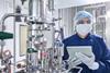
“When operating on a tumor, we can now use MS-based technologies to check if the surgical margins on tumor resection are clean and if all tumor cells have been removed,” says Heeren. How is this done? While the surgeon cuts with the diathermic knife, the smoke that comes from it is captured. By analyzing this smoke with mass spectrometry an overview of metabolic, lipid, or protein profiles per cell can be created. To check for the presence of tumor cells the profile of the smoke is compared with information from a molecular complexity model constructed from resected tumor tissue. The analysis of the smoke can be done in seconds.
“We do this for research purposes. The technique has already proven itself in the laboratory setting, but unfortunately, it is not yet an officially approved diagnostic device. It will take some time before the official authorities approve it.
In the meantime, we are working on reducing the size of the device so that it takes up less floor space in the OR and the noise level is also reduced.”
Near future
“In the near future, we hope that the work of the pathologist on material resected in the OR which now takes half an hour- making coupes, staining tissue, examination- can be done in one minute with our MSI. The MSI should then be able to scan and obtain information per cell at a rate of hundreds of thousands of cells in one minute.”
We are excited to welcome these speakers to Lab Technology 2024, at the Analytical Solutions conference. This conference will take place on May 28, 2024, in Reehorst, Ede.
You can register via this link.
Register now for participation in the conference and access to Lab Technology. See you May 28 in Ede!