
According to biomedical scientists at KU Leuven, the function of the digestive system appears to be much more sophisticated than simply pumping food around. In an article published in Nature, they demonstrate how the nervous system in mouse intestines distinguishes between different nutrients.
The intestinal wall is lined with around 100 million nerve cells, collectively known as the enteric nervous system. This system coordinates peristalsis, the wave-like contraction of muscles that pushes food forward. For a long time, researchers considered this to be a simple pumping system in which nerve cells communicated with each other in response to stretching or pressure. However, in recent years, more and more evidence has emerged suggesting that the system is more complex and sophisticated than previously thought.
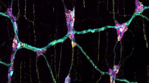
Biomedical scientists at KU Leuven, led by bioengineer Pieter Vanden Berghe, have now shown that the enteric nervous system responds not only to pressure, but also recognises different nutrients, such as sugars, proteins, and fats. To demonstrate this, the researchers used fluorescent markers that made the nerve cells glow bright green when activated. The researchers discovered that different subtypes of sensory nerve cell lit up in response to different types of nutrient. Vanden Berghe said, “Although we cannot yet explain the function of this with certainty, it is highly likely that the different neurochemical reactions accelerate or delay certain effects.”
Glowing cells
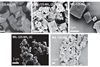
Vanden Berghe’s research group, LENS (Laboratory for Enteric NeuroScience), specialises in light microscopy for imaging living cells. They are among the few researchers in the world who use techniques in the laboratory that allow them to visualise both blood circulation and nerve cells in the intestines. They also perform intravital microscopy, which enables them to study the intestinal wall in living mice. To this end, they have developed computer algorithms that compensate for intestinal movement during image processing.
To visualise nerve cells, the researchers worked with transgenic mice. These mice were bred to carry calcium-sensitive green fluorescent protein (GFP) in their enteric nervous system. Vanden Berghe explains, “This meant that only the nervous system in the intestines was fluorescent. What’s more, the cells glowed even more intensely when activated by increased calcium.’
The researchers filmed small, round tissue samples of the intestinal wall while adding nutrients from above. They also used intravital microscopy to observe living mice. ‘By looking at the size of the cells, for example, we were able to distinguish different lighting patterns, where one subtype is larger than the other,’ says Vanden Berghe. ‘We also fixed pieces of tissue afterwards using immunohistochemistry, which allowed us to distinguish the different subpopulations because they each stain differently.’
The researchers also studied the connection between the different layers of nerve cells. “In most mammals, the intestines consist of two layers. The first layer is responsible for peristaltic mobility, while the second layer underneath controls blood flow. We observed that many sensory nerve cells in the first layer send signals back to the second layer.”
Meticulous work
According to Vanden Berghe, the intravital study was the biggest challenge. “It brought together so many things: biology, microscopy, and ethics.” During filming, the mouse remains anaesthetised. The mouse is centimetres in size, its intestine is millimetres, and with microscopy, we are looking at micrometres. Capturing that on camera was quite a task.”
A great deal of precision was also required to study the connection between the two layers of the intestine. However, postdoctoral researcher Candice Fung, the study’s first author, managed to achieve this, says Vanden Berghe. “To prove this, she conducted experiments on preparations that were 200 micrometres thick and 5 millimetres in diameter. She peeled away one layer, filmed the underlying cells, replaced the layer and then filmed the whole thing again. This showed that this connection was physically necessary for the cells to light up. When I show Candice’s work in presentations, everyone goes silent for a moment.”
Focus
The researchers now want to investigate how nerve cells respond to toxic or irritating substances. They also hope to focus on other locations in the intestine. “For practical reasons, we were only able to image a specific region.”
The researchers have now developed microscopic techniques that allow them to examine two areas of the intestine simultaneously. “We want to apply this in future research, together with nonlinear optical techniques.”


Nog geen opmerkingen