
Single particle cryo-EM offers the possibility of mapping proteins with atomic resolution. However, this technique is difficult and time-consuming because measurable samples require a large, homogeneous amount of the target protein. Now, structural biologists from Vrije Universiteit Brussel have presented a new microfluidic platform called MISO to Nature Methods. This reduces the necessary protein purification by up to a factor of one thousand.
Single-particle cryogenic electron microscopy (cryo-EM) is currently the preferred technique for measuring protein structures. By visualising thousands to a few million copies of a purified protein in a nanometre-thick layer of ice, the technique can image the protein in three dimensions with atomic precision.
In theory, only a few picograms of the protein are required, which can be purified from a single cell colony. In practice, however, it is not yet possible to purify proteins in such minimal, homogeneous quantities and place them directly into the sample holder. Instead, nearly a million times more biological material is required. This bottleneck means that determining the structure of certain proteins remains impossible.
However, structural biologists at the Vrije Universiteit Brussel (VUB) have now developed a new micro-isolation method that combines microfluidic protein purification with the preparation of the cryo-EM grid – the sample holder in the electron microscope. They validated their method by determining the structure of bacterial and eukaryotic membrane proteins using less than a microgram of protein. The transfer from cells to the cryo-EM grid took only a few hours.
Rouslan Efremov, a structural biologist at the VUB who led the research, explains: ‘Previously, researchers had to grow up to 150 Petri dishes full of cells to purify enough proteins for their structure determination. We demonstrate that MISO achieves the same result using only half a Petri dish of cells. ‘
Miniature method
For the current single-particle cryo-EM workflow, researchers need several milligrams of purified proteins — a million times more than they ultimately visualise. Due to this relatively large quantity, it is impossible to determine the structure of proteins from individual patient tissues. Efremov: ‘We wanted to simplify the preparation of the cryo-EM sample by bringing all the steps together on a microfluidic chip. ‘
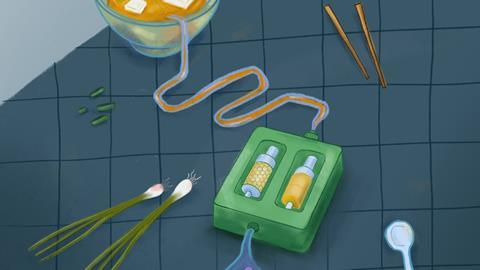
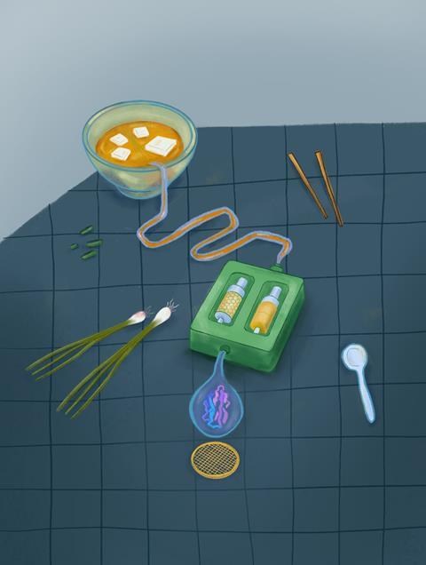
The result is MISO: a device that purifies proteins on a microfluidic chip and then loads them onto the cryo-EM grid via a capillary. ‘Researchers usually manually pipette a few microlitres of their purified protein onto this grid ‘, says Efremov. ‘But our chip can dispense a dozen nanolitres of the protein. ‘
Flow-through workflow
Instead of millilitres of buffer, the researchers needed only a dozen microlitres to determine the structure of several known membrane proteins. ‘Essentially, the purification process we use on our chip is the same as that used in standard setups ‘, says Efremov. ‘The difference is in the scale. Our purification columns are a thousand times smaller, which allows us to work with much smaller volumes. ‘
Although the idea of reducing and integrating all steps on a chip sounds simple, Efremov says it was very difficult to implement. ‘The big challenge was running the entire workflow on a chip in one go. In the lab, there are intermediate steps, such as keeping purified proteins in a test tube. But the volumes in a microfluidic chip are so small that you can ‘t just take them out and set them aside. It took a lot of work to make everything reproducible and reliable in one go, from cell extraction to loading into the cryo-EM grid. ‘
Wide application
The researchers now hope to expand the use of MISO. ‘Our current chip is made from the polymer PDMS. We are now looking to produce a prototype in large quantities from thermoplastics, which can then be used relatively cheaply in different laboratories. ‘
The researchers also want to determine how widely applicable MISO is. For example, they intend to use it to determine the structure of other proteins. The focus is mainly on proteins from human tissues. ‘Determining the structure of proteins from human, or even patient, tissues can provide insights into the influence of specific diseases or mutations. If we can visualise these low numbers thanks to MISO, it would be groundbreaking. ‘
Eluru, G. et al. (2025) Nat Methods, DOI: 10.1038/s41592-025-02894-x


Nog geen opmerkingen