


Avondlezing door prof.dr. E.M.J. Verpoorte georganiseerd door de Groningse Chemische Kring
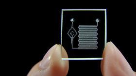
At the heart of microfluidics technology are planar devices based on networks of interconnected microchannels having sub-mL volumes. At these reduced dimensions, liquid transport is generally dominated by viscous forces, and flow streamlines are well defined and predictable. Thus, microfluidics offers a route to ultra-small-volume liquid handling for (bio)chemical applications with an unparalleled degree of control over liquid and particle handling. The first successful demonstrations of chip-based analysis in the early nineties involved the fast separation of fluorescent dyes and fluorescently labeled amino acids by capillary electrophoresis (CE) in microchannels several cm long. Since then, researchers from a wide range of disciplines have adopted this technology to perform not only analytical tasks, but synthetic chemical, biochemical, and biological tasks as well. Now a mature technology, microfluidics has enabled the miniaturization of established analytical techniques, thereby enhancing achievable performance especially with respect to reduction of analysis times. The ability to control solution transport in the nL to pL range so predictably has also been exploited to develop completely new approaches to conduct chemical and biological processing. Cells have dimensions in the micrometer range, and thus are easily confined and cultured in microfluidic channels and chambers to recreate microtissues with organ-like function. The resulting microsystems, referred to as organs-on-a-chip, have the special asset that they enable cellular microenvironments resembling those in the body to be created for these microtissues. This is accomplished by again exploiting the controlled flows typical of microfluidic devices, this time to establish carefully tailored concentration and shear stress gradients. The idea of interrogating physiological processes outside a complex living organism becomes realistic, with the generated data providing a more relevant reflection of that organism’s functioning. Microfluidics has thus been embraced especially by researchers in the life sciences seeking means to improve the quality of the physiological information they can obtain in their experiments with living tissue. This presentation will provide an overview of developments in microfluidics technology over the past 35 years since its inception (and the time that I have been privileged to be working in the field). I will illustrate this presentation with examples of work that I have been involved in at the University of Groningen.
More information can be found on the website of the lecture.

Venue
Sociaal Cultureel Centrum ‘t Clockhuys, Haren
Organiser
Groningse Chemische Kring