

With infrared spectroscopy in a mass spectrometer you can combine the information from both measurement techniques. If you compare the resulting IR spectrum with DFT calculations, you can unravel the precise molecular structure of biomarkers.
When screening for metabolic diseases and disorders, you look for specific biomarkers that could indicate a defective metabolic pathway in the body. In the metabolic disease pyridoxine-dependent epilepsy (PDE-ALDH7A1), the L-lysine pathway is interrupted by the malfunctioning of the enzyme α-aminoadipic semialdehyde dehydrogenase, also known as antiquitin. This causes the accumulation of α-aminoadipic semialdehyde (α-AASA), piperidine-6-carboxylate (P6C) and other substances, with P6C inhibiting pyridoxal-5’-phosphate and thus inhibiting the absorption of vitamin B6. The result is, among other things, epileptic seizures and, in many cases, a developmental defect of the brain. The earlier the diagnosis, the better the prognosis for people with this condition.
‘Instead of measuring IR and MS separately, we use infrared ion spectroscopy’
The problem is that the metabolites that are currently screened for are not stable. Therefore, researchers from Radboud University Medical Centre (Radboudumc) and Radboud University Nijmegen (RU) looked for other metabolites that could indicate PDE-ALDH7A1 in newborns. ‘The primary metabolites are known, but you can’t find them in a sample, so they are not suitable as biomarkers’, says Jos Oomens, professor of molecular structure and dynamics at the RU. ‘Our discovery is more concerned with an end product which does accumulate.’ That makes it possible to screen for those substances, and a technique has recently been developed to do just that: next-generation metabolic screening (NGMS).
‘NGMS was developed at the Radboudumc from the need to find new biomarkers for various metabolic diseases’, Oomens continues. ‘We are assigned the unknown substances.’ The Nijmegen professor’s team then uses a combination of tandem mass spectrometry (MS/MS) and infrared (IR) laser spectroscopy. ‘Instead of measuring IR and MS separately, we use infrared ion spectroscopy, also known as IRIS. The density of ions in a normal MS is too low to apply absorption spectroscopy to, and IR on its own cannot be used to identify individual components in a complex mixture.’
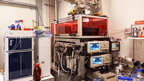
FELIX
The measurements are done with the free electron laser FELIX, which can be tuned to any desired IR wavelength. ‘We put a patient sample in the MS that ionises it and converts it to the gas form’, Oomens explains. ‘In the MS we isolate ions of the desired molecular weight and then emit the IR laser beam onto such a cloud of ions, giving us an IR spectrum linked to a specific mass.’
‘Because the metabolic pathway is known, the precursors are also known; that makes the puzzle a little easier’
Via NGMS, the colleagues at the Radboudumc found an increased concentration of an unknown substance with a mass of 186 Da in the lysine pathway: the first piece of the puzzle. ‘Via MS/MS we saw that the unknown substance breaks down to a mass of 128 Da, which corresponds to P6C, an important molecule in this pathway; we could confirm this with IRIS’, says Oomens. Such an IRIS spectrum can be compared with DFT calculations, which predict an IR spectrum of small molecules quite accurately (see picture below). But that was not all they needed; what is the molecule behind the mass of 186 Da?
After putting some of these pieces together, the mass difference seemed to suggest that P6C had an extra C3H5O group. Oomens: ‘Because the metabolic pathway is known, the precursors are also known. That made the puzzle a bit easier and it reduced the candidates to ten to twenty.’ After they had a match between the measured spectrum and the DFT calculations, it turned out that these were two diastereomers of (6-(2-oxopropyl)piperidine-2-carboxylic acid with almost the same retention time.
‘We then asked our synthesis colleagues in Floris Rutjes’ group and they made the two stereospecific molecules.’ With the two reference molecules Oomens and his group could then compare the spectra of the unknown biomarkers with the reference spectra, and thus definitively assign the molecular structures.
Screening of new-borns
The result is on the one hand practical because the university hospital could already use this method for the screening of newborns. ‘Just like the neonatal heel prick is already used to screen for other metabolic diseases’, Oomens explains. ‘Early detection of PDE means patients have a much better life expectancy, and you could start with an appropriate diet much earlier.’
On the other hand there is the academic result. ‘Now that we have identified these molecules, we can start answering the question: what is the pathophysiological effect of these metabolites? Could we find a potential treatment with these data?’ In addition, the picture of the metabolic pathway of lysine is now more complete. Oomens concludes: ’I find the cooperation between the hospital and the various university groups very valuable. Without cooperation you get nowhere.’
Van Outersterp, R.E. et al. (2021) Anal. Chem. 93(46)
Engelke, U.F.H. et al. (2021) J. Clin. Invest. 131(15)
The scope of IRIS
Infrared ion spectroscopy combines the information you can get from mass spectrometry (mass and elemental composition) and infrared spectroscopy (molecular structure). Although IRIS is still a fairly laborious technique, it can offer a solution when NMR measurements are difficult, for example because the concentration is too low. ‘This technique is just as sensitive as MS’, says Jos Oomens of Radboud University. ‘You may not be able to look at biopolymers with it, but small molecules with functional groups, such as metabolites and medicines, are very suitable for IRIS.’
Because looking at the metabolic pathways of endogenous substances is not the only thing you can use IRIS for. Together with Janssen Pharma, Oomens is looking at transformation products of medicines. Oomens: ‘Which molecules are created when the liver oxidises or breaks down a drug? You could also look at designer drugs, and find out if a methyl group has just shifted position, for instance in the case of the drug MMC. By putting the DFT spectrum over your measured spectrum, you can see the difference between 2-MMC and 3-MMC.’ Another application is to look at degradation products of aerosols such as α-pinene.
There are still some challenges. ‘If you don’t know what the molecule could be, for instance in the case of contaminants in water, it becomes a bit complicated’, Oomens explains. ‘The trick is to find a good workflow to identify the substances. You could think of machine learning or artificial intelligence, for instance. There are many compound libraries with millions of entries. You should be able to make a decent start with that.’
EuroFAST 2022
The FAST conference, which stands for Forum on Analytical Science and Technology, takes place every year. A few hundred analytical chemists from industry and academia attend. From 19-22 April 2022 the first European version - EuroFAST - will be held where Jos Oomens will also provide a lecture. ‘I want to tell about IRIS there and show new applications where possible’, he says. ’It is a good platform to make contacts, and as chairman of the Analytical Chemistry Section I find the insights from industry and universities a nice combination.’ Check out the website for more information and the possibility to register.