
Live visualisation of battle between cell and virus
With the new imaging technique VIRIM, you can follow the course of a virus infection in greater detail than ever before. A virus has even more tricks up its sleeve than expected.
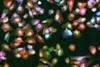
A small green dot under a microscope. When cell biologist Sanne Boersma spotted that point of light for the first time, she knew her experiment had succeeded. In other words: imaging technique SunTag - developed a few years earlier at the Hubrecht Institute in Utrecht, where Boersma works as a PhD student - can be used in viruses. The method brings ready-made fluorescent proteins to the scene of a virus infection: the translating and replicating viral RNA.
It means you can follow the first steps of a virus infection molecule by molecule, live and in living cells. It is the first time that researchers have been able to visualise this process in such detail. Boersma and colleagues called the application of SunTag VIRIM (virus infection real-time imaging) and published the accompanying paper in Cell.

Want to read more?
Create a free account today!
- Gain access to all our content on chemistry, life sciences and process technology;
- Get our weekly newsletter so you never miss a story.
As a member of the KNCV, KVCV, NBV, or NVBMB you have unlimited access. Log in here.